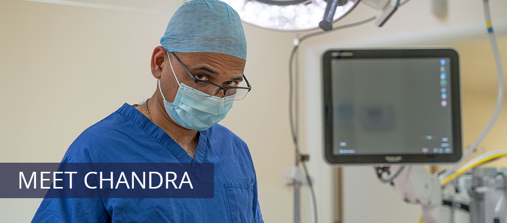
The information outlined below on common ankle conditions and treatments is provided as a guide only and it is not intended to be comprehensive. Discussion with Mr Chandra Rao is important to answer any questions that you may have.
For information about any additional conditions not featured within the site, please contact us for more information.
COMMON ANKLE CONDITIONS
Diagnosis
Standing X-rays are essential and some blood tests may be needed to check for rheumatoid arthritis. However advanced imaging is usually the key, especially computed tomography (CT) scans, or magnetic resonance imaging (MRI) or a bone scan (SPECT). In complex cases the precise injection of steroids under image guidance can give temporary relief and confirm the site of the damage. This will suggest the effectiveness of any permanent surgical treatment.
Treatment
The full benefit of non-operative treatments such as insoles, pads, shoes, physiotherapy and medicines will need to be assessed before embarking on surgery. An injection of steroids into the worn ankle joint can often provide excellent pain relief for some months.
Surgery
In early ankle arthritis, a catching pain, restricted movement or intermittent locking may be helped by minimally invasive technique of arthroscopic surgery. The gold standard technique has been fusion of the ankle (the two bones grow into one) where the painful grinding movements are eradicated. This is also performed using a minimally invasive technique.
Diagnosis
Our experts will listen carefully to your description of how your ankle is unstable. From this we can often determine if you problem is structural, functional or a combination of the two. Examining your ankle will allow us to assess if the ligaments are working properly and how strong the muscles controlling the ankle are. An MRI scan may be required both to look at the ligaments in more detail and to look for other problems such as cartilage damage, which may causing functional problems with your ankle.
Treatment
The first line of treatment for instability of any kind is usually physiotherapy. This strengthens and retrains the muscles stabilising the ankle, making it less prone to giving way. If your foot shape predisposes you to rolling your ankle we will arrange for you to have orthotics to support your foot. If your instability continues despite appropriate rehabilitation we may suggest surgery.
Surgery will be targeted towards the cause of your instability. The most common procedure is to tighten and reconstruct the damaged ligaments (a Broström-Gould procedure). We will also need to address any causes of pain which may be inhibiting your ankle control. This is usually done with a key-hole (arthroscopic) procedure at the time of your other surgery. You will normally need to be in some form of cast or removable boot for a month after surgery and then into a brace to protect the repair as you return to function. The hard work you put in before surgery with the physiotherapist will accelerate your post-operative recovery. This surgery is a reliable technique with a high success rate at returning athletes back to their pre-injury level of performance.
Diagnosis
Usually we can diagnose the problem by examining your leg. If the problem is at your heel we will arrange standing X-rays to look at the heel bone and the attachment of the tendon. An ultrasound or MRI can help quantify the problem, which is especially useful in troublesome cases or when considering more advanced treatments.
Treatment
The vast majority of people will get better with time, reducing their activity levels and careful calf stretches. We will arrange for a physiotherapist from our local team of experts to ensure you have the correct stretching technique. The eccentric stretches (lowering below a step) can be very effective but challenging and it important that you are doing them correctly. If you have insertional tendinopathy, a simple insole to raise your heel and ensuring that your shoe does not rub on the lump will help. If needed a more advanced orthotic will help to improve the forces going through the tendon.
If you are not responding after 3 months of stretching we will reassess and suggest which second line treatments would suit you. We frequently use shockwave therapy in such patients with good results. Occasionally we may recommend surgery if other treatments fail. Surgery is not without risk and can take a long time to fully get over so our surgeons will need to be happy that all appropriate other treatments have not worked.
COMMON ANKLE TREATMENTS
(1) arthritis of the joint, because of a previous injury that has damaged the joint, a generalised condition such as osteoarthritis or rheumatoid arthritis, or because the joint is just wearing out for some other reason
(2) severe deformity of the rear part of the foot, such as a flat foot, high-arched or “cavus” foot, a club foot or other deformity, in which the ankle joint is also deformed, unstable or damaged.
Why would it be done?
It is now possible to treat some arthritic ankle joints by replacing the joint, in the same way as arthritic hips and knees can be replaced. However, this is only suitable in older patients without major foot deformities, or people with rheumatoid arthritis or similar diseases. It would not be suitable if:
- you are young (usually under 45) or very physically active
- you have a severe foot deformity
- your ankle is very unstable
- you have had infection in the ankle or the bones around it
- the bone under the ankle (the talus) has collapsed
In these situations a fusion would be advised instead. If you have a severe foot deformity you may be advised to have this corrected at the same time as your ankle fusion by fusing other joints and/or breaking and realigning the foot bones. This would be discussed at the same time as your ankle fusion and we have other information sheets which give information about major foot fusions.
We are sometimes asked if a fusion can be changed to an ankle replacement later. This is not possible, as the foot becomes too stiff for an ankle replacement to work.
We often inject local anaesthetic or steroid into damaged joints, before any surgery is considered, to see whether this helps the pain. In some people, this gets rid of the pain and surgery is not necessary. In others, pain relief does not last but the results of the injection helps us to decide which joints to fuse.
What does it involve?
Ankle fusion in this hospital is nearly always performed by an arthroscopic (telescope) technique. This involves inserting a telescope into the ankle and by using specialised instruments we can remove the joint surface to allow the two bones to heal together. The bones are held rigidly by two screws inserted from the inner aspect of the leg just above the ankle joint. The operation will involve therefore 4 small cuts of approximately 1cm around the ankle.
Some people who have foot deformities have a tight Achilles tendon (“heel cord”), or weak muscles, or both. The Achilles tendon may be lengthened during surgery by making three small cuts in the calf and stretching the tendon.
What will happen after I go home?
By the time you go home you will have mastered walking on crutches without putting weight on your foot. You should go around like this for 2 weeks.
10-14 days after your operation you will be seen again in the clinic. Your plaster will be removed and the cuts and swelling on your foot checked. If all is well you will be put back in plaster or a brace. You should continue walking with your crutches but at this stage you can begin putting a little weight through your foot.
About 6 weeks after your operation you will come back to the clinic for an Xray. If this shows the joint is healing in a good position you can start putting about most of your weight through the plaster or brace. The physiotherapist will teach you how to do this.
You will have further Xrays once 3 months have elapsed. If the Xrays show that the joint is fused enough to take your weight, the plaster will be removed and you can start walking without it. Some people need to stay in plaster longer than 3 months.
An arthroscopy is a type of keyhole surgery used to diagnose and treat problems with joints.
Arthroscopy equipment is very small, so only small cuts in the skin are needed. This means it has some advantages over “open” surgery, including:
- less pain after the operation
- faster healing time
- lower risk of infection
- you can often go home the same day
- you may be able to return to normal activities more quickly
When an arthroscopy is used
You might need an arthroscopy if you have problems such as persistent joint pain, swelling or stiffness, and scans have not been able to find the cause. An arthroscopy can be used to assess the level of joint damage resulting from an injury, such as a sports injury, or from underlying conditions that can cause joint damage, such as osteoarthritis.
The procedure can also be used to treat a range of joint problems and conditions, including:
- repairing damaged cartilage
- removing fragments of loose bone or cartilage
- draining away any excess fluid
- treating arthritis
How an arthroscopy is carried out
Before having an arthroscopy, you’ll usually need to attend a pre-admission clinic. During the appointment, your general health will be assessed to make sure you’re ready for surgery. You’ll also be given information about issues such as:
- what and when you can eat and drink on the day of surgery
- whether you should stop or start any medicines before surgery
- how long it will take for you to recover from surgery
- whether you’ll need to do rehabilitation exercises after surgery
Your surgical team will explain the benefits and risks of having an arthroscopy. You’ll also be asked to sign a consent form to confirm you agree to have the operation and that you understand what’s involved, including the potential risks.
The procedure
An arthroscopy is usually carried out under general anaesthetic, although sometimes a spinal or local anaesthetic is used. Your anaesthetist will explain which type of anaesthetic is most suitable for you. Sometimes, you may be able to say which you would prefer.
If you have a local anaesthetic, your joint will be numbed so you do not feel any pain. You may still feel some sensations during the procedure, such as a slight tugging, as the surgeon works on the joint. Antibacterial fluid is used to clean the skin over the affected joint and a small cut, a few millimetres long, is made in the skin next to the joint so that an arthroscope (a thin, metal tube with a light and camera at one end) can be inserted. One or more additional incisions will also be made so that an examining probe or other fine surgical instruments can be inserted.
The joint is sometimes filled with a sterile fluid to expand it and make it easier for the surgeon to view. The arthroscope sends images to a video screen or eyepiece, allowing the surgeon to see inside your joint. As well as examining the inside of your joint, if necessary, your surgeon will be able to remove any unwanted tissue or repair damaged areas using tiny surgical instruments inserted through the additional incisions.
After the procedure, the arthroscope and any attachments are removed, along with any excess fluid from the joint. The incisions are usually closed using special tape or stitches and covered with a sterile dressing. An arthroscopy usually takes 30 minutes to 2 hours, depending on the type of procedure carried out. You’ll be able to go home on the same day as the surgery or the following morning.
Recovering from an arthroscopy
How long it takes to recover from an arthroscopy depends on the joint involved and the specific procedure you had. It may be possible to return to work within 7 days if your job involves sitting at a desk, but if it’s more physical, you may need to stay off work for up to 2 weeks. You may not be able to do more demanding physical activities, such as lifting and sport, for several months. Your surgeon or care team will let you know how long it’s likely to take to recover and what activities to avoid until you’ve fully recovered. While recovering, contact your GP or surgical team for advice if you think you may have a complication, such as an infection or blood clot.
Complications of arthroscopy
An arthroscopy is generally considered to be a safe procedure, but like all types of surgery there’s a risk of complications. It’s normal to have shortlived problems such as swelling, bruising, stiffness and discomfort after an arthroscopy. These usually improve in the days and weeks after the procedure. More serious problems are much less common (less than 1 in 100) and include:
- a blood clot that develops in a limb – known as deep vein thrombosis (DVT), it can cause pain and swelling in the affected limb
- infection inside the joint – known as septic arthritis, it can cause fever, pain and swelling in the joint
- bleeding inside the joint – which often causes severe pain and swelling
- accidental damage to the nerves near the joint – which can lead to temporary or permanent numbness and some loss of sensation
Speak to your surgeon about the possible risks before agreeing to have an arthroscopy.
Why would it be done?
They are frequently given for painful joints, trapped nerves and around ligaments. In the foot, these injections are often given to help in the diagnosis as well to control the symptoms. Some of these injections can be given in an outpatient clinic but others need to be given in an operating theatre using X-rays to guide the needle.
What does the injection involve?
The foot is placed in position so that the required joint can be seen on the X-ray and the local anaesthetic and steroid is injected in one go.
What will happen afterwards?
You can go home when comfortable and safe. It is advisable to rest your foot/ankle for the remainder of the day. The local anaesthetic usually wears off after 6 – 10 hours and your foot may feel bruised for a couple of days. You can take your normal painkillers if necessary. You can return to your normal activities as soon as you are able. The steroid takes 2 – 3 weeks to have is maximum effect and it may last for several months. You will be seen in outpatients 6 – 8 weeks after your injection.
Although hip and knee replacements are commonplace procedures, attempts to deliver the same solution for the ankle have been problematic. Devices have frequently loosened and failed, due to the pressure placed on the ankle. The ankle is a particularly complex joint to replace because it needs to flex and rotate and due to its size and shape, the ankle receives more strain than any other joint in the body. However the new generation of total ankle replacements integrate much more effectively into the ankle joint with much improved results.
This is a major procedure which is only considered for patients with advanced arthritis or severe damage to the joints following trauma. Joint wear and tear between the surfaces of the shin bone (tibia) and ankle bone (talus) will be extensive, causing pain even while resting and preventing normal daily activities.
The best results from total ankle replacement have been recorded in older patients, typically aged over 60, because they put their ankles under less stress than a younger person. Some younger patients have had good results from this type of surgery, but it remains to be seen how long the joint replacement lasts. A younger or particularly active person may consider an ankle fusion operation as a more appropriate alternative.
What does the operation involve?
Ankle replacement involves replacing the natural surfaces of the ankle joint which have degenerated with an artificial cover known as prosthesis.
The ankle replacement has three components. Two of the components cover the joint and in the middle there is a third, mobile component. This allows for greater movement and reduces the stress between the bone and the implants.
The component which covers the part of the ankle joint known as the tibia is flat. It is integrated into the bone with a short stem. The component which covers the part of the ankle joint known as the talus is curved and fixed into place with pegs. All the components are covered in a bioactive coating which encourages the patient’s own bone to grow into the artificial fixtures.
This type of operation allows the patient to preserve the movement that they have and gain a few degrees. Its success lies in the way that the ankle replacement integrates into the natural bone. The use of a mobile plastic bearing means the prosthesis is under much less stress and will stay in place a lot longer than the old two-part ankle joint replacements.
Recovery
A day after your surgery, you will be encouraged to take a few non-weight bearing steps with crutches under guidance of a physiotherapist. Your mobility on crutches and pain control will improve and you are likely to be discharged from hospital two to three days after surgery. You will be wearing a lightweight cast and walking confidently with crutches.
For the first six weeks after surgery, you will work with a physiotherapist on improving mobility and begin to partially weight bear. Your cast will be removed between four and six weeks following surgery, depending upon your recovery.
For the first six months following surgery, activities such as tennis and aerobics should be avoided. Six months post surgery, you should have normal joint mobility and be able to resume normal daily activities. However, it can take more than a year before you are fully recovered and the ankle replacement surgery can be completely evaluated.
Why would it be done?
Triple fusions are done for two main reasons:
(1) Arthritis of the joints, because of a previous injury that has damaged the joints, a generalised condition such as osteoarthritis or rheumatoid arthritis, or because the joint is just wearing out for some other reason
(2) Severe deformity of the foot, such as a flat foot, high-arched or “cavus” foot, a club foot or other deformity. Sometimes these can be corrected by breaking and reshaping the bones, but in other cases it is best to stiffen the joints in the corrected position, particularly if the joints are already stiff or the foot is weak.
If the damage or deformity is limited to one or two of the foot joints it may be possible to treat it by a more limited operation. However, as these joints work together, damage to one is often accompanied by damage to others.
We often inject local anaesthetic or steroid into damaged joints, before any surgery is considered, to see whether this helps the pain. In some people, this gets rid of the pain and surgery is not necessary. In others, pain relief does not last but the results of the injection helps us to decide which joints to fuse.
What does the operation involve?
Two cuts are made, one along the outer side of the foot and one on the inner side. Usually these are 4-5 cm long. Each of the three joints is opened up and the joint surfaces removed and, if necessary, reshaped to correct a deformity. The joints are then put in the correct place and fixed together with screws, pins or staples. The heel (“subtalar”) joint is usually fixed with a screw passed through a small cut in the back of the heel. The other joints are fixed through the main cuts.
It is usually necessary to put some extra bone into a triple fusion to get it to heal and to fill any gaps in the fusion left by correcting deformity. Often this extra bone can be obtained from the bone that is cut out to prepare the fusion. Sometimes there is not enough bone from this and bone has to be taken from the tibia bone just below the knee. Occasionally if a large amount of bone is required then this must be taken form the pelvis just above the hip.
Some people who have foot deformities have a tight Achilles tendon (“heel cord”) or weak muscles, or both. The Achilles tendon may be lengthened during surgery by making three small cuts in the calf and stretching the tendon. Some people with deformities of the foot also have deformed toes. Again, these may be corrected at the same time or at a later operation.
What happens after I go home?
By the time you go home you will have mastered walking on crutches without putting weight on your foot. You should go around like this for a month. 10-14 days after your operation you will be seen again in the clinic. Your plaster will be removed and the cuts and swelling on your foot checked. If all is well you will be put back in plaster. You should continue walking with your crutches.
About a 4-6 weeks after your operation you will come back to the clinic for an X ray. If this shows the joints are healing in a good position you can start putting most of your weight through the plaster. The physiotherapist will teach you how to do this
You will have further X rays once 3 months have elapsed. If the X rays show that the joint is fused enough to take your weight, the plaster will be removed and you can start walking without it. Some people need to stay in plaster longer than 3 months.
Achilles tendonitis is inflammation of the tendon at the heel or just above the heel. When non-surgical treatments like physiotherapy are unsuccessful, surgical intervention (key hole and/or open surgery) have an important role to play in giving much needed relief.
These injuries are common and can be debilitating. Surgery along with good rehabilitation is the key to early return to sports. Most surgeries are performed through minimally invasive or key hole procedures.
The information outlined above is provided as a guide only and it is not intended to be comprehensive. Discussion with Mr Chandra Rao is important to answer any questions that you may have.
QUICK ENQUIRY


CONTACT INFORMATION
Private Secretary: Jo Evans
joanne.evans66@nhs.net
NHS Secretary: Jo Brindle
jo.brindle@wvt.nhs.uk